
The spine is composed of 33 vertebrae. Seven of these bones are cervical vertebrae, 12 are thoracic vertebrae, and five are lumbar vertebrae. Five fused bones form the sacrum, and four fused bones form the coccyx, also known as the tailbone. The spine's function is to protect the spinal cord and provide support to keep the body in an upright position.
Except for the first and second cervical vertebrae and the fused vertebrae, there are supportive structures called discs between the other vertebrae. Discs consist of a soft, jelly-like center called the nucleus pulposus, surrounded by a tougher, elastic structure called the annulus fibrosus. Discs support the vertebrae and distribute the load placed on them, absorbing some of the shocks and providing flexibility.
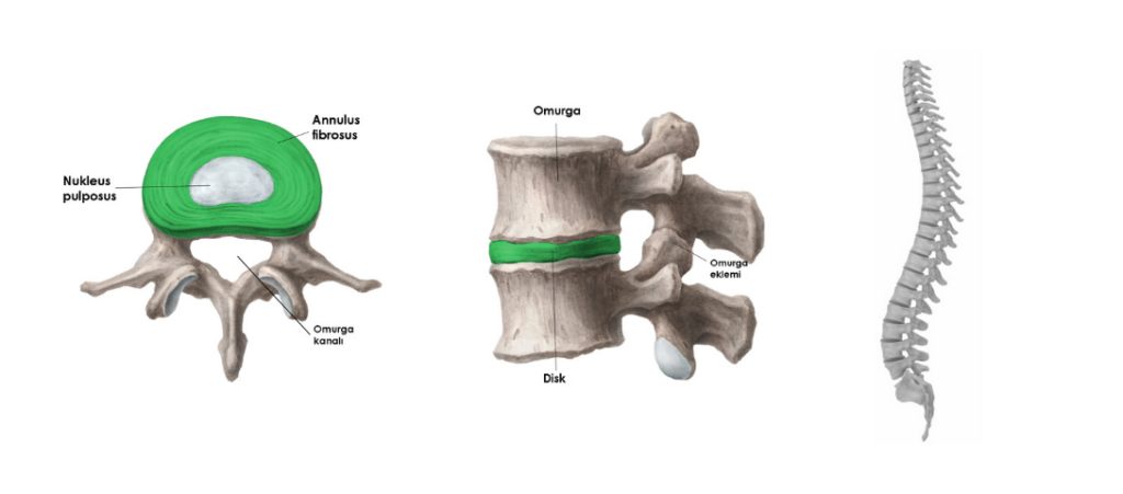
Vertebrae are connected to each other by disc structures, ligaments, and small joint structures. When the vertebrae are connected in this way to form the spine, the small canal behind each vertebra combines to form a cylindrical canal called the spinal canal. The spinal cord is located within this canal. It extends from the first cervical vertebra (C1) to the first lumbar vertebra (L1) as a continuation of the brainstem. The remaining section consists of nerve roots resembling a horse's tail (cauda equina). Thirty-one pairs of spinal nerves, forming the peripheral nervous system, emerge from the spinal cord, with eight pairs in the cervical region, 12 pairs in the thoracic region, five pairs in the lumbar region, and six pairs in the sacral region. These nerves supply the arms, trunk, legs, and internal organs.