
The nervous system is generally divided into the central nervous system and the peripheral nervous system. The central nervous system consists of the brain, brainstem, cerebellum, and spinal cord. The peripheral nervous system consists of the spinal nerves emerging from the spinal cord and the autonomic nervous system.
The brain and spinal cord are located within a bony structure called the cranium and the spinal canal, which protect them from impacts. The brain and spinal cord are surrounded by membranes called meninges. The meninges consist of three layers. From outer to inner: dura mater, arachnoid, and pia mater. The dura mater is thick, whitish, and inflexible. Below this, there is a thinner, transparent-looking part called the arachnoid. Beneath this is the pia mater, a very thin layer that surrounds all the convolutions of the brain and spinal cord. There is a subdural space between the dura and arachnoid and a subarachnoid space between the arachnoid and pia.
The brain is divided into two main parts: the right and left hemispheres, which are connected by special structures at specific locations. The internal structure of the brain consists of gray and white matter. Gray matter forms the cortex, which is composed of many nerve cells (neurons) about 2-5 mm thick. This structure is a convoluted structure consisting of many indentations and swellings. Beneath this is the white matter, which consists of connection fibers between neurons.
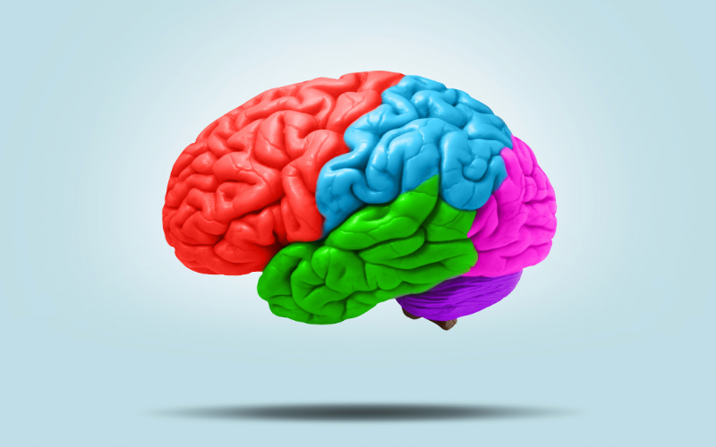
The right and left hemispheres of the brain are divided into specific regions (frontal, parietal, temporal, occipital lobes) based on various characteristics. Each region has specialized centers that perform specific functions and are interconnected (such as the speech center, comprehension center, auditory center, visual center, etc.).
Within the brain, there are small cavities called ventricles that are connected by thin channels. These cavities are filled with cerebrospinal fluid produced by specialized structures called choroid plexus. The cerebrospinal fluid flows through these channels to the subarachnoid space surrounding the brain and spinal cord and is absorbed into the venous system from there.