The spine consists of 33 vertebrae. Seven of these bones are cervical vertebrae, 12 are thoracic vertebrae, and 5 are lumbar vertebrae. Five of them are fused to form the sacrum, and 4 are fused to form the coccyx, also known as the tailbone. The function of the spine is to protect the spinal cord and provide support to keep the body in an upright position.
Except for the first and second cervical vertebrae and the fused vertebrae, there are supportive structures called intervertebral discs between adjacent vertebrae. Intervertebral discs consist of a soft, jelly-like center called the nucleus pulposus surrounded by a tougher outer layer called the annulus fibrosus. Discs support the vertebrae, distribute the load on them, and absorb some of the shocks, acting as flexible structures.

The vertebrae are connected to each other by disc structures, ligaments, and small joint structures. The interconnected vertebrae form a cylindrical channel called the spinal canal by joining the small channels located at the back of each vertebra. The spinal cord is located within this canal, extending from the first cervical vertebra (C1) to the first lumbar vertebra (L1) as a continuation of the brainstem. The section beyond this point is composed of nerve fibers in a horse's tail-like arrangement (cauda equina). Thirty-one pairs of spinal nerves, forming the peripheral nervous system, emerge from the spinal cord as pairs on each vertebral level. Eight of these are cervical nerves, 12 are thoracic nerves, 5 are lumbar nerves, and 6 are sacral nerves. These nerves extend to the arms, trunk, legs, and internal organs.

Piriformis syndrome is a neuromuscular condition that occurs when the piriformis muscle, located deep within the hip region, compresses or irritates the sciatic nerve passing nearby. While this muscle performs a critical function by enabling the external rotation of the leg, it exerts pressure on the sciatic nerve—the longest nerve in the body—in cases of […]
View in Detail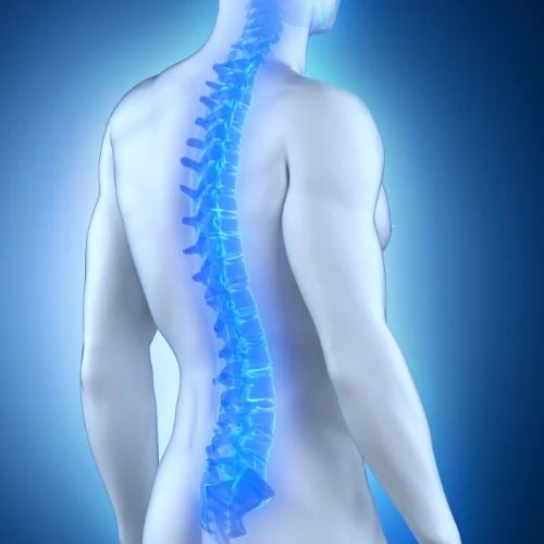
Basic Anatomy of The Spine and Spinal Cord The spine consists of 33 vertebrae. Seven of these bones are cervical vertebrae, 12 are thoracic vertebrae, and 5 are lumbar vertebrae. Five of them are fused to form the sacrum, and 4 are fused to form the coccyx, also known as the tailbone. The function of […]
View in Detail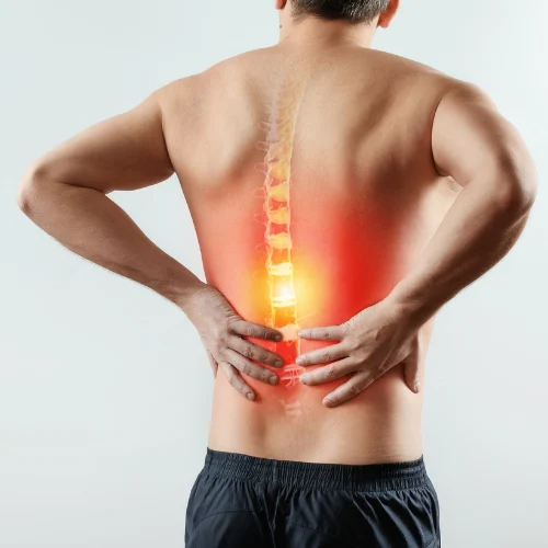
Disc Herniation Herniated discs in the neck (cervical), back (lumbar), or more rarely in the mid-back (thoracic) are illness caused by degeneration of structures called discs located between the spinal bones, due to various reasons such as trauma, heavy lifting, excess weight, aging, poor posture, and genetic predisposition. Following the degeneration of the annulus of […]
View in Detail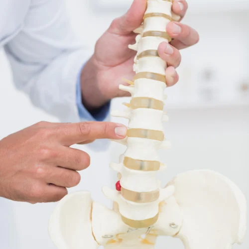
Spondylolisthesis (Spinal Slippage) Spondylolisthesis, or spinal displacement (spinal slippage), is the displacement of one or more vertebrae in the spine that are normally aligned, due to various reasons. This displacement can occur forwards, backwards, to the right, or to the left, affecting the neck, back, or lower back. It can occur in a single vertebra […]
View in Detail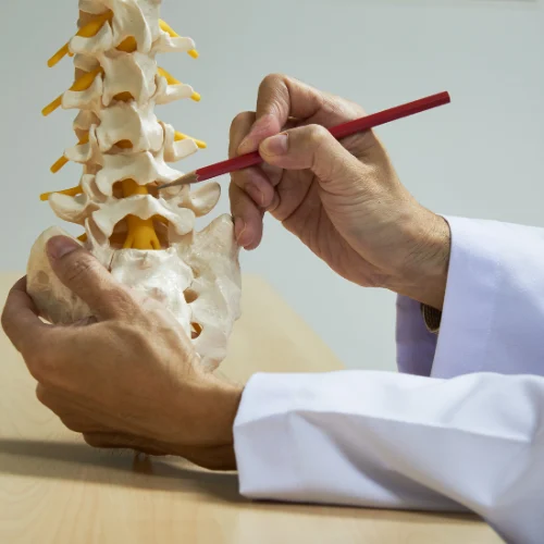
Spinal Stenosis (Narrowing of The Spinal Canal) Spinal stenosis, commonly known as "narrow canal," is a condition characterized by the narrowing of the spinal canal for various reasons, leading to pressure on the spinal cord and the nerves passing through it. This narrowing can occur at any level of the spine, including the neck (cervical), […]
View in Detail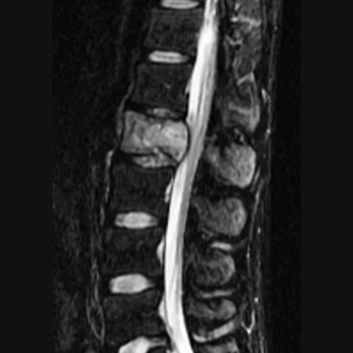
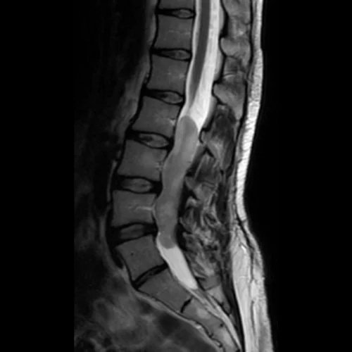
Spinal Tumors: Symptoms, Diagnostic Methods, and Current Treatment Approaches Spinal tumors are abnormal masses of cells that develop within the spinal canal or the bone structures (vertebrae) that make up the spine. These tumors can originate directly from the spine's own tissues (primary tumors) or occur when cancer from another part of the body (such […]
View in Detail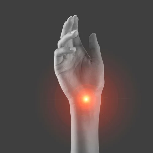
Carpal Tunnel Syndrome Carpal tunnel syndrome is a condition that occurs when the median nerve, one of the major nerves in the hand, is compressed and subjected to pressure within a structure called the carpal tunnel at the wrist level, due to various reasons. Causes include repetitive hand and wrist movements (computer use, playing musical […]
View in Detail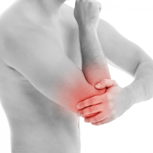
Cubital Tunnel Syndrome Cubital tunnel syndrome is a condition that occurs when the ulnar nerve, one of the three main nerves in the hand, becomes compressed or exposed to pressure at the elbow region. The nerve travels between two bony protrusions on the inner side of our elbow and passes through a structure called the […]
View in Detail
Thoracic outlet syndrome (TOS) is a complex condition that occurs as a result of the compression of nerves or blood vessels passing through the narrow space between the clavicle (collarbone) and the first rib, known as the thoracic outlet. This narrowing typically results from traumatic injuries, repetitive arm and shoulder movements, postural disorders, or anatomical […]
View in Detail
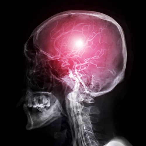
Basic Anatomy of Central Nervous System The nervous system is generally divided into the central nervous system and the peripheral nervous system. The central nervous system consists of the brain, brainstem, cerebellum, and spinal cord. The peripheral nervous system consists of spinal nerves originating from the spinal cord and the autonomic nervous system. MENINGES (BRAIN […]
View in Detail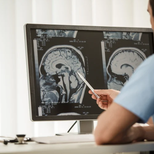
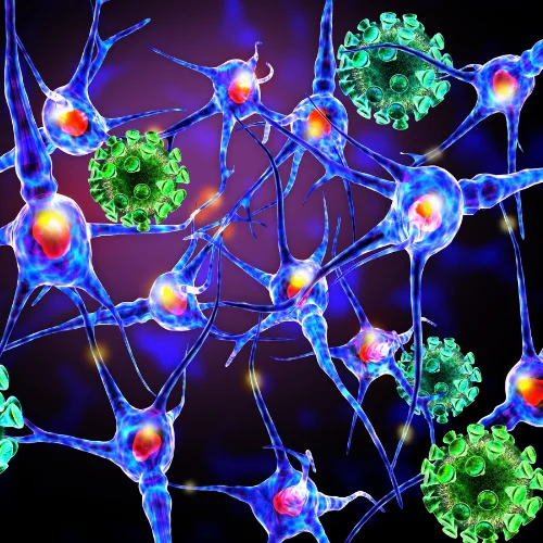
Brain Tumors The causes of brain tumors are not fully understood, but risk factors include radiation exposure, genetic disorders, a family history of tumors, diseases that affect the immune system, stress, and exposure to various environmental carcinogens. It is thought that brain tumors occur as a result of damage or improper function of specific genes […]
View in Detail
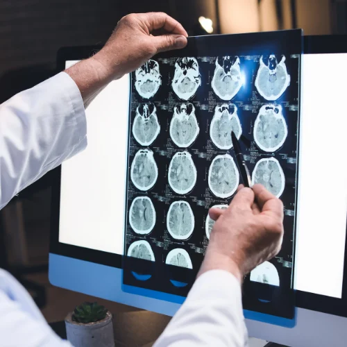
Pituitary Tumor The pituitary gland is a vital endocrine gland located at the base of the brain in a small bony cavity called the "sella turcica" (Turkish saddle) and is widely known as the body's "conductor of the orchestra." Pituitary tumors are masses that arise from the abnormal and uncontrolled growth of cells within this […]
View in Detail
Chiari malformation (commonly known as Chiari syndrome) is a structural disorder characterized by the protrusion (herniation) of cerebellar tissue through the opening at the base of the skull (foramen magnum) into the spinal canal. Under normal conditions, the parts of the cerebellum should be located entirely within the skull; however, they are pushed downward due […]
View in Detail